









What Do Healthy Dog Gums Look Like? Colors and Red Flags
Healthy dog gums are smooth, moist, and salmon-pink to bubblegum-pink on unpigmented areas. They look intact, and a gentle press turns a pink patch white before the color returns within about 1–2 seconds.
If the gums are pink, is the mouth healthy? Most owners stop there, and that is the gap. A textbook shade ignores this dog. The useful comparison is this dog's own usual color, moisture, and refill speed, not the dog next door.
What do healthy dog gums look like on a quiet afternoon is a screening question, not a clearance. A tidy lip line can sit over disease in the pocket around the tooth. A calm look in good light tells you whether today matches the mouth you already know. It doesn't measure what you can't see.
Table of Contents
- Healthy Dog Gum Color, Texture and Surface at a Glance
- Pigmented and Spotted Gums That Can Be Normal
- How to Check Your Dog's Gums at Home
- Build a Gum Baseline Log to Track Change
- Reading Capillary Refill Time Results
- Abnormal Gum Colors, Signals and Causes
- Healthy-Looking Mouths Can Still Hide Dental Disease
- Know When to Call the Vet About Gums
Healthy Dog Gum Color, Texture and Surface at a Glance
On unpigmented tissue, healthy gums are smooth, moist, and salmon-pink to bubblegum-pink, and the surface stays intact, with no swelling, sores, or bleeding that starts on its own. Merck Veterinary Manual's overview of periodontal disease treats that look as a screening observation. The benchmark is consistency with this dog's usual appearance, not a neighbor's mouth.
Six features carry the check. Color, texture, surface, refill, temperature, and pigment each answer a different question. Missing one of them is how a "looks fine" mouth gets filed away too early.
| Feature | Healthy appearance | Why it matters |
|---|---|---|
| Color | Salmon to bubblegum pink on unpigmented areas | Pink tissue suggests adequate blood flow; compare with the dog's usual color |
| Texture and moisture | Smooth and moist | Dry or tacky gums can signal dehydration or illness |
| Surface | Intact, with no ulcers, bleeding, or swelling | Sores, bleeding, or swelling point to gum disease, trauma, or infection |
| Capillary refill time | Pink returns in about 1–2 seconds after pressure | Slower refill can mean poor circulation |
| Temperature | Warm, similar to the rest of the body | Cold gums can signal poor circulation |
| Pigmentation | Brown, black, or mottled areas can be normal | Judge new or changing pigment against the dog's own stable pattern |
What each feature is telling you
Color on a pink patch is a blood-flow clue. A dog that has always been a softer salmon isn't "wrong" next to a brighter bubblegum shade. Texture and moisture should feel slick, not sticky. Dry gums often show up with illness before the color shifts much.
Surface integrity is the part owners rush past. Intact means no ulcers, no spontaneous bleeding, and no puffy margin. Bright red, swollen, bleeding, ulcerated, or painful tissue isn't a normal variant. It can reflect gingivitis, trauma, infection, or systemic disease.
Capillary refill time is the press test. On a pink area, the tissue blanches white and returns in about 1–2 seconds. That window is a quick read on peripheral circulation, not a lab result. Temperature should feel warm, close to the rest of the body. Cold gums belong with a circulation concern, not with a cosmetic note.
Pigmentation varies by dog. Brown, black, or mottled patches can be normal and longstanding. You judge them against that dog's stable pattern.
Practical rule: Match what you see to the table, name the feature that's off, and don't treat a pretty surface as proof the mouth is clear.
These checks spot change. They don't replace a veterinary oral exam, because disease can sit below the gumline while the visible surface still looks relatively normal. If color, moisture, surface, refill, temperature, or pigment doesn't match the table, move to a structured check or to the abnormal-color patterns below.
Pigmented and Spotted Gums That Can Be Normal
A healthy mouth doesn't have to be uniformly pink. Plenty of dogs carry brown, black, or mottled pigment, and that tissue can be entirely ordinary. Owners swing both ways. They treat lifelong dark gums as disease, or they file a new spot under "just pigment" and stop looking.
PetMD's guide to dog gum colors draws the line that actually helps. Pigment that is longstanding, flat, and stable is a different finding from pigment that is new, enlarging, raised, bleeding, or tied to a lump. Color alone isn't the test. Healthy tissue is generally moist. Bleeding, marked redness, swelling, pain, foul odor, or a new growth needs a veterinary look.
Stable versus concerning
Stable pigment usually shares these traits:
- Older photos match it: The same patch shows up months ago, in the same place.
- It lies flat: No thickening, no raised edge, no change in how the lip sits over it.
- It's quiet: Moist, painless, and unchanged in size, often roughly symmetrical if both sides have always been dark.
Concerning change looks different, even on a dog whose mouth has always been spotted:
- New or growing: A spot that wasn't in last month's photo, or one that's thicker than it was.
- Surface trouble: Raised texture, bleeding, ulceration, odor, pain when the lip is lifted, or a lump beside the pigment.
Inspect the entire gumline, including the tissue over the back molars, not only the incisors people see when the dog pants. Compare today's mouth with older photographs of the same spots. A dark patch you can't assess for color or refill isn't a failed check. Move to a normally pink section for those reads, and log the dark areas on their own.
Change over time and the signs that travel with it matter more than any universal pink standard.
Keep the check visual, calm, and well lit. Don't scrape, pick, or press a suspicious spot to "see what it does." Manipulation can bleed a lesion and muddy what the vet needs to see. Anything new, raised, enlarging, or painful gets an exam rather than a home experiment.
How to Check Your Dog's Gums at Home
Bright light does more for this check than a stronger grip. Dim rooms make pink tissue look pale, and a phone flash washes the shade you came to judge.
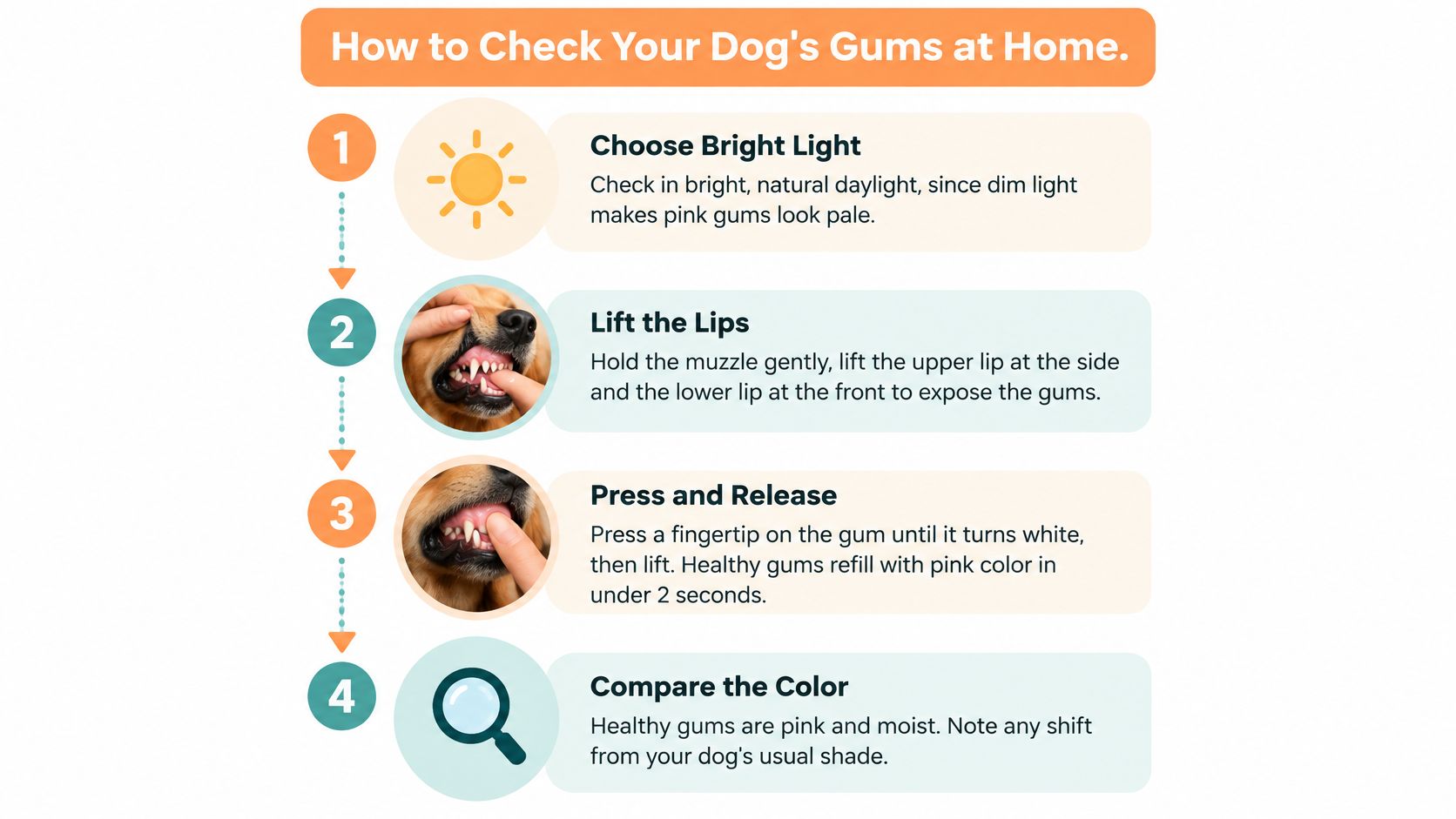
The sequence
- Pick daylight and a calm moment. Artificial dimness shifts pink toward pale, so a window or an outdoor porch beats a hallway lamp.
- Hold the muzzle gently and lift the lips. Raise the upper lip at the side and the lower lip at the front so you can see gum above the canine and the incisors without prying the jaw open.
- Find a pink, unpigmented patch. Note that shade against this dog's usual look. Dark pigment won't give you a fair color or refill read.
- Press, release, and count. Use a fingertip until the spot turns white, then lift. Healthy pink returns in about 1–2 seconds. One calm press beats three hard ones.
- Scan past the front teeth. Follow the gumline to the back molars. Front-only checks miss redness, tartar, and lumps owners never see in a smile.
- Read the rest of the surface. Moisture, bleeding, swelling, sores, odor, and lumps belong in the same pass as color.
- Photograph that same pink patch. Same light, same distance, saved to a dated log. A monthly repeat is what makes slow change visible.
Stop if the dog pulls away or stiffens. A bitten finger isn't a better data point. Never poke, scrape, or pick at a spot to test it.
Slips that spoil the reading
Pressing hard or repeating the press on the same spot distorts refill. Rushing the count turns "about two seconds" into a guess. Shade and flash both lie about color. If you want a fair baseline, do this once in daylight and keep that setup.
A short walk-through of the lip lift and the press helps the first time you try it.
If the scan shows tartar along the gumline, color isn't the only home task. A separate routine for removing tartar from dog teeth at home covers what brushing and wipes can and can't reach. Gum color still doesn't clear the tooth roots.
Build a Gum Baseline Log to Track Change
Start the log the first time you get a calm, well-lit look, before anything seems wrong. Later checks only help if the first entry is already on the phone.
Repeat monthly, or the same day something looks different. I ask owners to judge the new photo against the first entry, not yesterday's. Slow drift hides when you only compare neighbors.
What goes in each entry
- Photograph one pink patch. Use the upper gum above the canine, same daylight and roughly the same distance. Flash shifts pink toward white or orange, so daylight stays the rule for every row.
- Write the refill in seconds. Three gentle presses on that same patch, then note the times. A single rushed count isn't a baseline.
- Describe the tissue in words. Color against this dog's usual look, whether it's moist, and any swelling, bleeding, odor, or lumps.
- Log dark patches on their own line. Size and position for each one, so a new spot can't hide inside "he's always had black gums."
- Compare with entry one. A stable pattern and a refill still in the normal window supports watching. A shift in color, refill, or shape is the reason to book, not to add another month of photos.
Bring the log to the visit and say the date the change first showed up. "It looks a bit pale" is weaker than a photo from March next to a photo from June. The vet still examines the mouth. Your log only shortens the history.
Reading Capillary Refill Time Results
Capillary refill time is a quick perfusion screen, not a diagnosis. On a visible pink area, press until the gum blanches, release, and watch the pink return. The Merck Veterinary Manual triage table lists approximately 1–2 seconds as the normal reference range.
Use the same pink patch each time, and repeat two or three gentle presses. Pigmented gum won't give you a readable blanch.
| Refill time | What it may mean | Next step |
|---|---|---|
| Under 1 second | Can occur in fever, heatstroke, or early shock, a hyperdynamic state | Check temperature, breathing, and energy; call the vet if the dog seems unwell |
| 1 to 2 seconds | Normal reference range | Save as your baseline and recheck if the dog changes |
| Over 2 seconds | Possible poor perfusion or peripheral vasoconstriction | Call the vet promptly; go now if gums are also pale or blue, or the dog is weak |
| No clear reading | Pigment, cold surroundings, stress, or technique can distort the test | Retest on a pink patch in good light, then ask the vet |
Bands, not verdicts
A refill under 1 second can show up in hyperdynamic states such as fever, heatstroke, distributive shock, or early compensatory hypovolemic shock. Fast isn't automatically "extra healthy." Pair it with how the dog is breathing and whether the body feels hot.
Over 2 seconds may reflect poor perfusion or peripheral vasoconstriction. That single number still isn't a cause. It tells you circulation to that patch is sluggish right now.
What throws the test off
Pigmentation, ambient temperature, technique, anxiety, and shifting circulation all change the count. A cold room, a nervous dog, or a press on a black patch can fake a delay. Read refill next to breathing rate and effort, temperature, pulse quality, energy, and hydration. One odd second with a bright, comfortable dog is a retest. The same second with other changes is not.
Delayed refill plus pale, white, or blue-gray gums, weakness, cold extremities, respiratory difficulty, collapse, bleeding, or suspected poisoning is an emergency. Don't finish the photo log first.
Abnormal Gum Colors, Signals and Causes
Abnormal color is a screening signal, not a diagnosis. A dog with a color change or signs of systemic illness needs prompt veterinary care. Dental products alone don't treat shock, anemia, or poor oxygenation. Veterinary Partner's mucous-membrane guidance is blunt on that point. Pale or white gums may track with anemia, blood loss, or reduced perfusion. Blue-gray can mean inadequate oxygenation. Bright red or markedly yellow membranes are abnormal too, especially with weakness, breathing changes, collapse, vomiting, or suspected toxin exposure.
Black pigment still can't be scored for color or refill. Use a normally pink section.
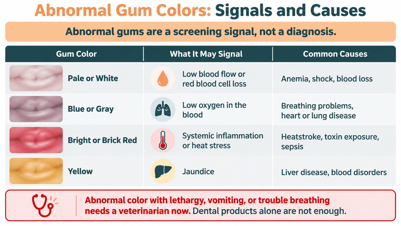
| Gum appearance | What it may signal |
|---|---|
| Pale pink to white | Anemia, blood loss, shock, or reduced blood flow |
| Blue, gray, or purple | Inadequate oxygen reaching tissues from heart, lung, or airway problems |
| Bright or brick red | Gingivitis, heatstroke, or toxin exposure |
| Markedly yellow | Possible liver, blood, or toxin problem |
| Pinpoint red or purple spots (petechiae) | Bleeding under the surface, such as low platelets or a clotting disorder |
| Swollen, bleeding, or ulcerated tissue | Gum disease, trauma, infection, or systemic illness |
Patterns by body system
Blood and circulation. Anemia, ongoing or internal blood loss, trauma, shock, and severe dehydration can all drain the pink out of a gum. In good light the tissue looks washed out, sometimes white, and it may feel cool or tacky rather than slick.
Oxygenation. Heart, lung, and airway disease can push gums toward blue, gray, or purple when tissues aren't getting enough oxygen. That shade with hard breathing isn't a lighting problem.
Oral disease. Gingivitis and periodontal disease often paint the margin bright red, swollen, or bloody. The visible rim can be only part of the problem, which is why a red line still needs an oral exam rather than a stronger toothpaste.
Heat and toxins. Heatstroke can flush gums bright or brick red. Some poisons, including certain rodenticides, show up as bleeding rather than a single neat color. Markedly yellow membranes raise concern for liver or blood disorders, or for toxin exposure.
Clotting. Pinpoint red or purple spots are blood under the surface, the sort of pattern seen with low platelets or a clotting disorder. They are small. You miss them if you only glance at the canine teeth.
How to describe the change
Compare the shade with your baseline photo. Note whether it appeared in an hour or over weeks. Then line it up with breathing, energy, appetite, vomiting, and anything the dog might have eaten or chewed. Bright red or yellow tissue becomes more urgent when weakness, breathing changes, collapse, vomiting, or toxin exposure sit beside it. Write those companions down before you call. The color word alone is a weaker history than color plus what else changed today.
Healthy-Looking Mouths Can Still Hide Dental Disease
Could your dog have dental disease even when the gums look pink and intact? Yes. Gum color and a refill test show the visible surface and offer a screening check, but they cannot show what is happening below the gumline. Compare your dog's mouth with its usual appearance and track changes over time. A healthy-looking margin is not an all-clear.
Periodontal disease develops in pockets around tooth roots, below the gumline, where a lip-lift cannot reach. Mild disease may be present without obvious outward signs. Only a veterinary oral exam, with probing and dental radiographs, can assess the tissues beneath the surface.
Signs that still count when the color looks ordinary
- Breath that stays foul after a drink or short walk, not only after meals.
- Tartar along the gumline, particularly on the back teeth.
- Loose or missing teeth, or a tooth that has shifted to a new angle.
- Reluctance to chew, dropping food, or chewing mostly on one side.
- Pawing at the mouth or pulling away when you lift a lip.
- Blood on a hard chew or toy, even if the gums looked pink earlier.
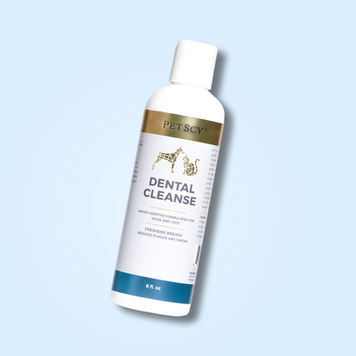
Home care helps manage reachable tooth surfaces between veterinary visits. Brushing, dental wipes, and water additives can support daily cleaning, but they do not measure pocket depth or reveal disease beneath the gumline. A dog dental hygiene routine that you can maintain week after week can complement, not replace, a veterinary exam. Keep watching for changes from your dog's normal mouth, even when the color appears ordinary.
Know When to Call the Vet About Gums
How quickly should you act when your dog's gums change? Match the response to the finding, and compare the color with your dog's usual baseline. A new change matters even if the gums do not look dramatic.
| Finding | How urgent | What to do |
|---|---|---|
| Pale, white, blue, or gray gums | Emergency | Go to an emergency vet now |
| Slow refill with weakness, collapse, or labored breathing | Emergency | Go now, even if gum color looks near normal |
| Suspected toxin exposure with any gum change | Emergency | Call a poison helpline and a vet immediately |
| Refill over 2 seconds without other signs | Prompt | Call your vet and arrange advice or assessment within 24 hours |
| Bleeding, ulcers, or swelling without other signs | Urgent | Arrange a vet visit within 24 hours |
| Pinpoint red or purple spots | Urgent | Arrange a vet visit within 24 hours |
| New, raised, or enlarging dark spot or lump | Soon | Book an exam within a week; sooner if it bleeds |
| Red gumline, tartar, or bad breath only | Routine | Schedule a dental exam |
| Stable pigment and normal refill in the photo log | Monitor | Recheck at routine visits |
Urgency isn't only the dramatic row
A single refill reading over 2 seconds warrants a prompt call, even if your dog seems well. Check the result once more under calm conditions, then contact your vet within 24 hours for advice. If weakness, collapse, or labored breathing appears, treat it as an emergency and go now.
Bleeding, ulcers, swelling, or pinpoint spots also need veterinary advice within 24 hours. A new, raised, or enlarging dark spot or lump can wait for an exam within a week only if it is not bleeding and your dog is otherwise well. Arrange care sooner if it bleeds or your dog will not eat.
A red gumline, tartar, or bad breath alone usually calls for a routine dental visit, not an emergency trip. Do not ignore persistent odor, though. If breath is the main concern, these practical steps for dog bad breath may help while you arrange an exam.
A stable pattern in your photo log and a refill within the normal window support monitoring at routine visits. Keep checking against your dog's own baseline, since pigment and color vary between dogs. Home checks cannot assess tissue beneath the gumline. A 2024 study in Scientific Reports assessed 127 client-owned dogs and classified only 5 dogs (3.9%) as having healthy gingiva under detailed periodontal assessment. A mouth can look fine at the lip and still need a professional examination.
Bring your photo log, note when the change began, and ask for a periodontal exam. Those details help your vet assess what has changed and decide what to check next.
PetScy offers veterinarian-formulated dental care for dogs, including options aimed at plaque, tartar, and breath. Products can support a home routine, but they cannot replace a vet assessment when gum color, refill, or comfort changes. Visit PetScy to see the dental range and choose an option that fits your routine.